
Download
Application Note
Expansion and cellular characterization of primary human adherent cells in the Quantum® Cell Expansion System, a hollow-fiber bioreactor system
Boah Vang, Nathan Frank, Mark Jones, Brian Nankervis, Claire Coeshott*
Terumo BCT, Inc., 10810 West Collins Avenue, Lakewood, CO 80215, USA
*Corresponding author: Claire Coeshott, Email: claire.coeshott@terumobct.com
Competing interests: The authors were all employees of Terumo BCT, Inc. at the time of this study.
Abbreviations used: A-MSCs, adipose-derived mesenchymal stem cells; BM-MSCs, bone-marrow-derived mesenchymal stem/stromal cells; CPPT, cryoprecipitate; EC, extra-capillary; FN, fibronectin; GMP, good manufacturing practice; IC, intra-capillary; LDH, lactate dehydrogenase; LN, laminin; PACs, primary adherent cells; PBS, phosphate-buffered saline; SkMMs, skeletal muscle myoblasts; TCPSs, tissue culture polystyrene surfaces
Received February 14, 2020; Revision received March 19, 2020; Accepted March 24, 2020; Published April 8, 2020
Abstract
Primary adherent cell types can be expanded in the Quantum® Cell Expansion System (Quantum system), an automated platform that utilizes a hollow-fiber bioreactor. This system can replace manual cell culture and produce cells that retain their phenotypes and functionality. Bone-marrow-derived and adipose-derived mesenchymal stem/stromal cells have previously been successfully expanded on the Quantum system. We have now successfully used the Quantum system to expand fibroblasts and myoblasts. Hollow-fiber bioreactors were coated with adherence-supporting proteins, and then cells were loaded and expanded in the appropriate growth medium for 7 to 15 d. Cells were harvested from the bioreactors using enzymatic reagents. Harvested cell yields ranged from 100 × 106 to 1 × 10⁹ cells, with viability typically above 90%. The number of doublings obtained from Quantum system harvests ranged from 4 to 9. The Quantum system is a functionally closed expansion system that can reduce contamination due to minimal interventions and can automate the culture process to reduce labor and reagent costs.
Keywords: adherent cells, fibroblasts, mesenchymal stem/stromal cells, myoblasts, Quantum cell expansion system
INTRODUCTION
Advances in regenerative medicine have shown that tissues and organs that are impaired by disease, injury, or age can be repaired using cellular therapy [1]. Primary adherent cells (PACs) such as bone-marrow-derived mesenchymal stem cells (BM-MSCs, also known as mesenchymal stromal cells or medicinal signaling cells) and adipose-derived mesenchymal stem cells (A-MSCs) have multipotent properties that make them ideal candidates in tissue regeneration [2]. Fibroblasts derived from primary somatic cells are also versatile adherent cells that play an integral role in the support and repair of tissues and organs due to their adaptability to respond to damage [3]. Skeletal muscle myoblasts (SkMMs) are another source of PACs that comprise renewable progenitor cells that participate in injury repair [4]. These PACs—BM-MSCs, A-MSCs, fibroblasts, and SkMMs—all have the potential to undergo differentiation into functional cells that contribute to structural regeneration of tissues.
However, the large numbers of ex vivo expanded cells that are required in many clinical cell therapy protocols make standard culture conditions (manual growth in low-volume cell culture flasks or plates) problematic and expensive because of the need for extensive personnel and facility resources and the potential for contamination [5]. To meet such clinical demand, a robust, automated, and closed cell expansion method is optimal. The Quantum® Cell Expansion System (Quantum system) (Terumo BCT, Inc., Lakewood, CO) is a functionally closed, automated hollow-fiber bioreactor system designed to reproducibly expand adherent or suspension cells in either good manufacturing practice (GMP) or research laboratory environments [6]. The Quantum system has dual-loop fluid pathways: the intra-capillary (IC) and extra-capillary (EC) sides of the semipermeable hollow fibers that allow for efficient nutrient and gas exchange and waste removal. Details of the Quantum system’s features and its advantages over manual cell culture processes have been highlighted in a recent review [7].
Original efforts to characterize cells grown in the Quantum system compared the genetic stability of BM-MSCs expanded on the Quantum system to those expanded on tissue culture polystyrene surfaces (TCPSs) [8]. Growth kinetics, trilineage differentiation potential, immunophenotyping, spectral karyotyping, in vitro micronuclei formation, and tumorigenesis in an athymic (nu/nu) mouse model were evaluated. The results indicated that BM-MSCs expanded on the Quantum system displayed similar characteristics to BM-MSCs expanded on TCPSs and that genetic stability was not affected [8]. Additionally, a comparison of manufacturing A-MSCs on the Quantum system versus manual processing in tissue culture flasks found that expansion rate and yield were significantly enhanced in the Quantum system and the purity and quality of cells were maintained [9].
We have now evaluated the use of the Quantum system with other adherent cell types and demonstrate here that the Quantum system can be successfully used for ex vivo expansion of human fibroblasts and SkMMs.
MATERIALS AND METHODS
Expansion and harvest of cells in the Quantum system
Unless otherwise noted, experiments on the Quantum system were performed as follows and as previously described in detail [6]. Disposable cell expansion sets containing hollow-fiber bioreactors were loaded onto the Quantum system and primed with phosphate-buffered saline (PBS) without Ca2+ and Mg2+ (Lonza, Walkersville, MD), using the pre-configured Prime Cell Expansion Set task to remove air from the set. Hollow fibers were coated overnight with 5 mg research-grade human fibronectin (FN) (Corning, Corning, NY) reconstituted with 10 ml distilled water and further diluted with 90 ml PBS. Pooled human cryoprecipitate (CPPT) (Vitalant, Denver, CO) diluted into single-donor equivalents with PBS was also used as an alternative to FN in fibroblast experiments. For SkMMs, bioreactors were coated overnight either with 5 mg FN or with 5 mg FN plus 1 mg human laminin (LN) (Sigma-Aldrich, St. Louis, MO), all diluted with PBS. Quantum system media bags were filled with growth medium components using peristaltic pumps. Cells and reagents being loaded onto the Quantum system were transferred into Quantum system cell inlet bags using syringes with luer lock tips. Auxiliary bags containing media, reagents, and cells were sterile-welded onto the Quantum system using a TSCD®-Q sterile tubing welder (Terumo BCT, Inc., Lakewood, CO). Media exchanges and bioreactor conditioning to appropriate gas concentrations were performed according to the operator’s manual. Gas mixtures of 20% O2, 5% CO2, balance N2 were attached to the Quantum system and the system maintained the temperature at 37°C.
To add cells to the IC side of the Quantum system, the automated task Load Cells With Uniform Suspension, described in the operator’s manual, was used. A customized cell loading procedure consisting of a series of sequentially decreasing circulation rates alternating the flow in the positive and negative directions was also used to load cells in some experiments. Glucose and lactate were monitored with an i-STAT 1 handheld blood analyzer (Abbott Point of Care, Princeton, NJ) to track cell metabolism. IC inlet rates were increased accordingly depending on metabolite levels. In general, if glucose levels were lower than 70 mg/dl, the IC inlet rate was doubled. Cells were expanded for 7 to 15 d, depending on the cell numbers loaded and on metabolic activity. Cell harvests were performed with the Release Adherent Cells And Harvest task described in the operator’s manual using trypsin (0.25% with EDTA) (Thermo Fisher Scientific, Waltham, MA) for fibroblasts and either trypsin or 1 mg/ml research-grade collagenase (Sigma-Aldrich) for SkMMs.
Cell count, viability, number of doublings
Cell counts were measured with a Vi-CELL™ XR automated cell counter (Vi-CELL, Beckman Coulter®, Indianapolis, IN) using trypan blue dye exclusion to assess viability. The number of doublings was calculated using the formula [LN(number of cells harvested/number of cells loaded)]/[LN(2)].
Fibroblasts
Several experiments on the Quantum system were completed comparing FN- and CPPT-coated bioreactors with precultured human dermal fibroblasts (Lonza Group Ltd, Basel, Switzerland (Lonza)). Fibroblasts were loaded into the Quantum system and cultured for between 7 and 11 d. Complete growth medium consisted of Clonetics FGM-2 BulletKit (Lonza) supplemented with 10% fetal bovine serum (FBS; (Thermo Fisher Scientific)). Complete growth media for paired runs with FN- and CPPT-coated bioreactor runs consisted of Iscove’s Modified Dulbecco’s Medium (Lonza) supplemented with 10% FBS and GlutaMAX.
Characterization of fibroblasts by phenotype
Phenotypic characterizations were evaluated on representative cell harvests from FN- and CPPT-coated runs by flow cytometry using antibodies with the following specificities: CD34, CD45, CD90 (all BD BioSciences, San Jose, CA) and fibroblast surface protein (FSP, Bioss, Woburn, MA). Cell staining was performed per the manufacturer’s instructions. Cell samples were acquired on a BD FACSCantoTM II (BD BioSciences) and analyzed with BD FACSDivaTM software.
Fibroblast potency assay
A Human Collagen Type I ELISA Kit (MD Biosciences, Oakdale, MN) was used to evaluate collagen production from harvested cells. The assay was carried out per the manufacturer’s instructions. Fibroblasts harvested from the Quantum system were digested for 72 h prior to assay. A standard curve was generated and concentrations for Quantum system samples were derived from the curve using their absorbance values.
SkMMs
Clonetics human skeletal muscle myoblasts (Lonza) were initially expanded in 525 cm2 cell culture multi-flasks (T525) with a starting seeding density of 1.5 × 10³ cells/cm2 and were maintained in a humidified tissue culture incubator at 37°C and 5% CO2. Cells were cultured in complete growth medium consisting of α-MEM with GlutaMAX (Thermo Fisher Scientific), 10% FBS (GE), 0.01 µg/ml epidermal growth factor (Thermo Fisher Scientific), and 4 µg/ml dexamethasone (Sigma-Aldrich). Once cells reached 80% confluence, they were washed with PBS, released with trypsin, and neutralized with complete growth medium. T525 cell harvests from replicate flasks were pooled, centrifuged, and re-suspended in complete growth medium. Cell counts and viabilities were obtained and SkMMs were subsequently loaded into the coated bioreactors of the Quantum systems and cultured between 7 and 15 d in complete growth medium. Following harvest, a lactate dehydrogenase (LDH) assay was performed on several runs to evaluate residual SkMMs in the bioreactors. LDH was measured with a Cytotoxicity Detection kit (Sigma-Aldrich) according to the vendor’s instructions.
Characterization of SkMMs by differentiation
SkMMs harvested from flasks or the Quantum system were plated in tissue culture plates, cultured in complete growth medium until approximately 80% confluent, then differentiated into myotubules as previously described [10].
RESULTS
Fibroblasts
Cell harvests from each experiment were analyzed to obtain the general growth characteristics (Table 1). Morphological observations, flow cytometry, and type I collagen assays were performed on representative samples. A range of 2 × 10⁶ to 20 × 106 cells were loaded into bioreactors to examine whether different seeding densities could lead to cell proliferation. The harvest yields ranged from 300 × 106 to 1 × 10⁹ total cells with all viabilities above 97%. There were no observable differences in growth kinetics among all the runs. An average of 6.2 ± 1.4 doublings (n = 16) was observed. Two different media formulations were used to test the Quantum system’s ability to expand fibroblasts in different conditions.
Fibroblasts expanded on the Quantum system maintained their fibroblastic morphology as displayed in Figure 1A-1D. The cells attached to their substrate and maintained bipolar or multipolar morphology with elongated shapes [11].
Surface marker expression profiles of Quantum-system-expanded fibroblasts had less than 1.4% expression for the negative markers CD34 and CD45 and more than 89% expression for the positive markers CD90 and FSP (Table 2). There was no difference between FN and CPPT (n = 3, Table 2).
Collagen type I was present in cells expanded on the Quantum system, indicating that they were collagen-producing fibroblasts. Cells harvested from FN-coated bioreactors (n = 6) compared to CPPT-coated bioreactors (n = 6) had similar average concentrations of collagen type I, 0.5 ± 0.5 µg/ml.
Table 1. Summary of growth characteristics for fibroblast expansions on the Quantum system.
| No. of Quantum runs (n) | No. of cells loaded (106) | Coating reagent | Culture time (d) | Individual No. of cells harvested (106) | Average No. of cells harvesteda (106) | No. of doublingsa | Viabilitya (%) |
|---|---|---|---|---|---|---|---|
| 2 | 2 | FN | 11 | 476 | 646 | 8.3 | 98.5 |
| 816 | |||||||
| 2 | 2 | FN | 10 | 1000 | 915 | 8.9 | 98.9 |
| 829 | |||||||
| 3 | 8 | FN | 8 | 395 | 348 | 5.4 | 99.0 |
| 347 | |||||||
| 302 | |||||||
| 2 | 17 | FN | 7 | 863 | 885 | 5.7 | 99.6 |
| 906 | |||||||
| 2 | 20 | FN | 7 | 863 | 902 | 5.5 | 98.0 |
| 940 | |||||||
| 3 | 8 | CPPT | 8 | 398 | 341 | 5.4 | 98.0 |
| 328 | |||||||
| 296 | |||||||
| 2 | 10 | CPPT | 9 | 384 | 380 | 5.3 | 99.0 |
| 376 |
aThe last three columns in the table indicate averages for the set of runs within each experiment. Coefficients of variation were < 10% for number of doublings and < 0.5% for viability.
Table 2. Phenotypic characterization of fibroblasts by flow cytometry (n = 3).
| Biomarker | FN-coateda | CPPT-coateda |
|---|---|---|
| CD90 | 97.5 ± 0.1 | 96.9 ± 0.2 |
| FSP | 88.9 ± 7.7 | 93.1 ± 5.1 |
| CD34 | 0.3 ± 0.1 | 0.5 ± 0.0 |
| CD45 | 0.91± 0.5 | 1.4 ± 0.8 |
aAverages and standard deviations using FN- and CPPT-coated bioreactors are shown for biomarkers with values displayed as percentage of cell population with positive expression.
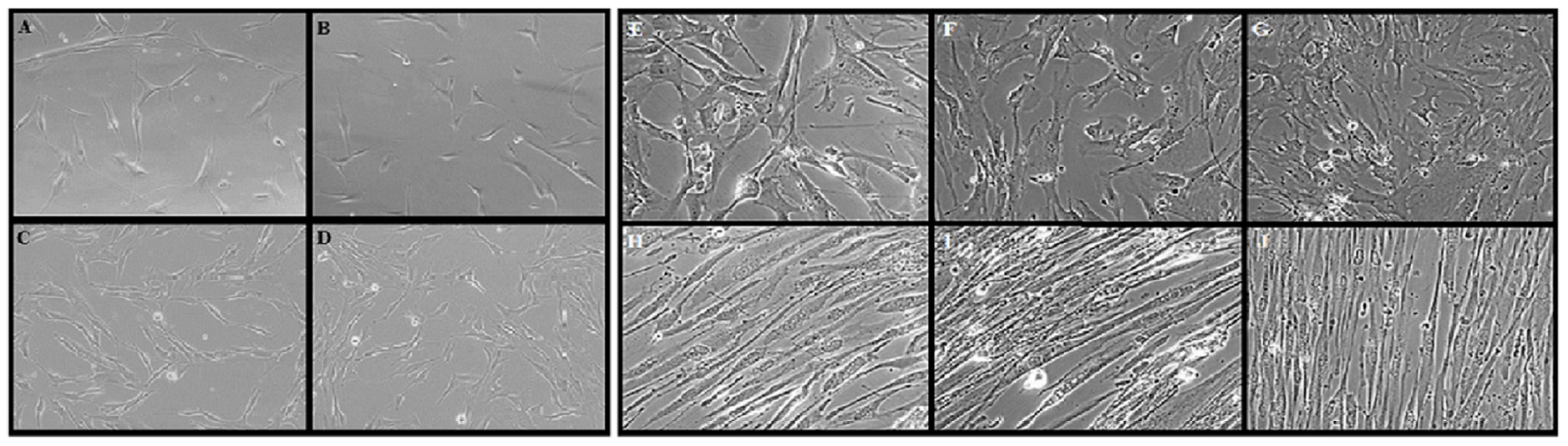
Figure 1. Morphology of adherent cells after expansion and harvest. Images were captured using an Olympus CKX41 inverted phase-contrast microscope at 100× magnification. A-D. Representative fibroblast harvests from Quantum system runs. Post Quantum system-harvested cells were seeded into 24 well cell culture plates and observed for morphological characteristics. Cells were from experiments in the following rows in Table 1: (A) row 5; (B) row 2; (C) row 3; and (D) row 6. E-J. SkMMs previously expanded in flasks or Quantum systems were plated in tissue culture plates, cultured in complete growth medium until approximately 80% confluent, then differentiated into myotubes. Non-differentiated SkMMs from flask-expanded cultures (E) and Quantum system-expanded cultures (F and G). Differentiated SkMMs from flask-expanded cells (H) and Quantum system-expanded cells (I and J).
Table 3. Summary of growth characteristics for SkMM expansions on the Quantum system.
| No. of cells loaded (106) | Coating reagent | Harvest reagent | Culture time (d) | No. of cells harvested (106) | Residual cellsa (106) | No. of doublings | Viability (%) | Average No. of cells harvested (106) |
|---|---|---|---|---|---|---|---|---|
| 2.0 | FN | Collagenase | 11.9 | 107 | 34 | 5.7 | 89 | 140 |
| FN | 14.9 | 172 | 65 | 6.4 | 97 | |||
| 4.8 | FN + LN | Trypsin | 9.9 | 238 | ND | 5.6 | 92 | 231 |
| FN + LN | 12.9 | 224 | 106 | 5.5 | 89 | |||
| 8.1 | FN + LN | Trypsin | 8.7 | 152 | 144 | 4.2 | 93 | 136 |
| FN + LN | 119 | 167 | 3.9 | 93 | ||||
| FN + LN | 138 | 129 | 4.1 | 93 | ||||
| 10.0 | FN | Collagenase | 6.9 | 394 | 46 | 5.3 | 97 | 576 |
| FN | 6.9 | 487 | 85 | 5.6 | 98 | |||
| FN | 6.9 | 403 | 77 | 5.3 | 97 | |||
| FN | 10.0 | 1020 | 138 | 6.7 | 98 |
aResidual cells estimated from LDH assay, not included in other growth characteristic calculations; ND: not determined.
SkMMs
Cells harvested from the Quantum systems were analyzed to obtain growth characteristics for SkMM expansions (Table 3). A range of 2 × 10⁶ to 10 × 106 precultured SkMMs were loaded onto bioreactors to examine whether different seeding densities could lead to cell proliferation. The harvest yields ranged from 107 × 106 to 1 × 10⁹ total cells with the highest cell loading number (10 × 106) achieving the highest cell yields. The LDH assay estimated that an average of 99 × 106 (± 45 × 106) cells, or an average of approximately 30% of total cells in the bioreactor, remained on the bioreactors after the initial harvest. An average of 5.3 ± 0.9 doublings (n = 11) was observed. Viabilities on all the harvested products ranged from 89% to 98%.
Other variables tested to evaluate preferred conditions for SkMM growth were the coating reagent, release agent, and culture time. There were no obvious differences in SkMM growth kinetics between FN- and (FN + LN)-coated bioreactors. The runs that were harvested with collagenase typically had fewer cells remaining on the bioreactors compared to the runs harvested with trypsin.
SkMMs harvested from two Quantum system-expanded runs were subsequently differentiated in parallel with cells that were only expanded in flasks. Representative microscopic images of randomly selected locations of each well were taken before and after differentiation to demonstrate morphological changes (Figure 1E-1J). Prior to differentiation, cells were widely spread with no apparent polarity. After 8 d of differentiation, cells became elongated with some polarity and branching [12]. Therefore flask-expanded and Quantum system-expanded SkMMs displayed similar morphological characteristics before and after induced differentiation. The capacity for myoblasts to fuse with each other to form myotubes is an important cellular function [13] and was exhibited in cells from these experiments.
DISCUSSION
We show here that two primary adherent cell types, fibroblasts and SkMMs, can be expanded in the Quantum system and we describe conditions for their growth. The Quantum system provides an automated and functionally closed system for cell culture that minimizes manual manipulations and open steps, thereby reducing labor and decreasing chances of contamination. Cellular characterization assays demonstrated that there were no significant changes in phenotype in cells expanded in the Quantum system compared to either flask-grown controls or literature references. The number of doublings that fibroblasts and SkMMs underwent on the Quantum system were on average 6.2 ± 1.4 and 5.3 ± 0.8, respectively. This translates to an average 130-fold and 45-fold increase in cell yield for fibroblasts and SkMMs, respectively, with some runs starting with as few as 2 × 10⁶ cells in the inoculum, highlighting the versatility of the Quantum system to support robust growth and expansion of a range of cell inocula. Different media formulations and bioreactor coating materials were also used on the Quantum system runs and were able to sustain cell expansion.
The expansion of non-adherent cells such as T cells has also been recently demonstrated in the Quantum system, which has potential for benefits in the cancer immunotherapy space [14]. Additionally, Russell et al. [15] recently demonstrated that BM-MSCs manufactured on the Quantum system are physically and functionally equivalent to BM-MSCs manufactured by manual flask methods and use of the Quantum system allowed significant cost savings per dose and manufacturing time.
The current studies were undertaken to assess the ability of the Quantum system to support expansion of diverse PAC types, and thus high cell yield was not the focus. Nevertheless, yields of up to one billion cells per Quantum system were obtained for both cell types. Results demonstrate that robust ex vivo expansion of cells to numbers sufficient for clinical cell therapy protocols is possible using the Quantum system. This opens the door for future applications for the Quantum system to expand cells for therapeutic use.
Acknowledgments
Thank you to the following former associates of Terumo BCT for their contributions to this article: Michelle Brecheisen, Domicinda Hill, Kim Nguyen, Rebecca Peters, and Thomas Startz.
References
- Mao AS, Mooney DJ (2015) Regenerative medicine: Current therapies and future directions. Proc Natl Acad Sci U S A 112: 14452-14459. doi: 10.1073/pnas.1508520112. [View Article] [PubMed] [Google Scholar]
- Ballas CB, Zielske SP, Gerson SL (2002) Adult bone marrow stem cells for cell and gene therapies: implications for greater use. J Cell Biochem Suppl 38: 20-28. doi: 10.1002/jcb.10127. [View Article] [PubMed] [Google Scholar]
- Alberts B, Johnson A, Lewis J, Raff M, Roberts K, et al. (2002) Fibroblasts and their transformations: The connective-tissue cell family. In: Molecular Biology of the Cell. 4th edition. New York: Garland Science. Available from: https://www.ncbi.nlm.nih.gov/books/NBK26889.
- Durrani S, Konoplyannikov M, Ashraf M, Haider KH (2010) Skeletal myoblasts for cardiac repair. Regen Med 5: 919-932. doi: 10.2217/rme.10.65. [View Article] [PubMed] [Google Scholar]
- Choi W, Kwon S, Jin HJ, Jeong SY, Choi SJ, et al. (2017) Optimization of culture conditions for rapid clinical-scale expansion of human umbilical cord blood-derived mesenchymal stem cells. Clin Transl Med 6: 38. doi: 10.1186/s40169-017-0168-z. [View Article] [PubMed] [Google Scholar]
- Hanley PJ, Mei Z, Durett AG, Cabreira-Harrison MdG, Klis M, et al. (2014) Efficient manufacturing of therapeutic mesenchymal stromal cells with the use of the Quantum Cell Expansion System. Cytotherapy 16: 1048-1058. doi: 10.1016/j.jcyt.2014.01.417. [View Article] [PubMed] [Google Scholar]
- Barckhausen C, Rice B, Baila S, Sensebé L, Schrezenmeier H, et al. (2016) GMP-compliant expansion of clinical-grade human mesenchymal stromal/stem cells using a closed hollow fiber bioreactor. Methods Mol Biol 1416: 389-412. doi: 10.1007/978-1-4939-3584-0_23. [View Article] [PubMed] [Google Scholar]
- Jones M, Varella-Garcia M, Skokan M, Bryce S, Schowinsky J, et al. (2013) Genetic stability of bone marrow-derived human mesenchymal stromal cells in the Quantum System. Cytotherapy 15: 1323-1339. doi: 10.1016/j.jcyt.2013.05.024. [View Article] [PubMed] [Google Scholar]
- Haack-Sørensen M, Follin B, Juhl M, Brorsen SK, Søndergaard RH, et al. (2016) Culture expansion of adipose derived stromal cells. A closed automated Quantum Cell Expansion System compared with manual flask-based culture. J Transl Med 14: 319. doi: 10.1186/s12967-016-1080-9. [View Article] [PubMed] [Google Scholar]
- Jarocha D, Stangel-Wojcikiewicz K, Basta A, Majka M (2014) Efficient myoblast expansion for regenerative medicine use. Int J Mol Med 34: 83-91. doi: 10.3892/ijmm.2014.1763. [View Article] [PubMed] [Google Scholar]
- Del Pino A, Ligero G, López MB, Navarro H, Carrillo JA, et al. (2014) Morphology, cell viability, karyotype, expression of surface markers and plasticity of three human primary cell line cultures before and after the cryostorage in LN2 and GN2. Cryobiology 70: 1-8. doi: 10.1016/j.cryobiol.2014.10.011. [View Article] [PubMed] [Google Scholar]
- Berendse M, Grounds MD, Lloyd CM (2003) Myoblast structure affects subsequent skeletal myotube morphology and sarcomere assembly. Exp Cell Res 291: 435-450. doi: 10.1016/j.yexcr.2003.07.004. [View Article] [PubMed] [Google Scholar]
- Nehlin JO, Just M, Rustan AC, Gaster M (2011) Human myotubes from myoblast cultures undergoing senescence exhibit defects in glucose and lipid metabolism. Biogerontology 12: 349-365. doi: 10.1007/s10522-011-9336-5. [View Article] [PubMed] [Google Scholar]
- Coeshott C, Vang B, Jones M, Nankervis B (2019) Large-scale expansion and characterization of CD3+ T-cells in the Quantum® Cell Expansion System. J Transl Med 17: 258. doi: 10.1186/s12967-019-2001-5. [View Article] [PubMed] [Google Scholar]
- Russell AL, Lefavor RC, Zubair AC (2018) Characterization and cost-benefit analysis of automated bioreactor-expanded mesenchymal stem cells for clinical applications. Transfusion 58: 2374-2382. doi: 10.1111/trf.14805. [View Article] [PubMed] [Google Scholar]